


An 8-year-old male was brought to the paediatric dermatology clinic with a sudden onset of target-like skin lesions on his extremities, face, and trunk. The lesions were preceded by an oral herpes outbreak. The child also reported fever, malaise, arthralgia, conjunctivitis, and oral ulcers. The patient complained of pain, itching, and difficulty swallowing due to oral involvement (Figures 1, 2). Physical examination revealed widespread erythematous and violaceous target lesions with central clearing, a hallmark of erythema multiforme. The patient’s mucous membranes, including the oral cavity and conjunctiva, were involved. The patient experienced painful urination due to inflammation of the urethral lining. Laboratory investigations showed mild leucocytosis and elevated inflammatory markers. The clinical presentation and compatible history led to the diagnosis of erythema multiforme major (EMM). HSV IgM and HSV IgG in the serum were positive, and mucosal involvement was seen, indicating that HSV-associated erythema multiforme (EM) due to herpes simplex virus (HSV) infection was the cause. The patient was prescribed 500 mg of azithromycin and 400 mg of acyclovir. The administration of acyclovir was maintained upon the detection of higher titre levels for HSV-1. Cyclosporine was introduced at a dose of 5 mg/kg of body weight. The patient showed significant improvement within the first week of treatment, with a decrease in the number and severity of skin lesions. Mucous membrane involvement also resolved, and the patient reported a reduction in pain and discomfort.
Figure 1
A – EMM commonly involves the oral mucosa, presenting as painful erosions or ulcerations. In our patient’s case, the lips, tongue, and buccal mucosa were affected. The eyelids were swollen due to fluid accumulation, with redness of the conjunctiva, but no damage to the cornea. B – Lesions with a distinctive target-like appearance located on the patient’s ear lobe
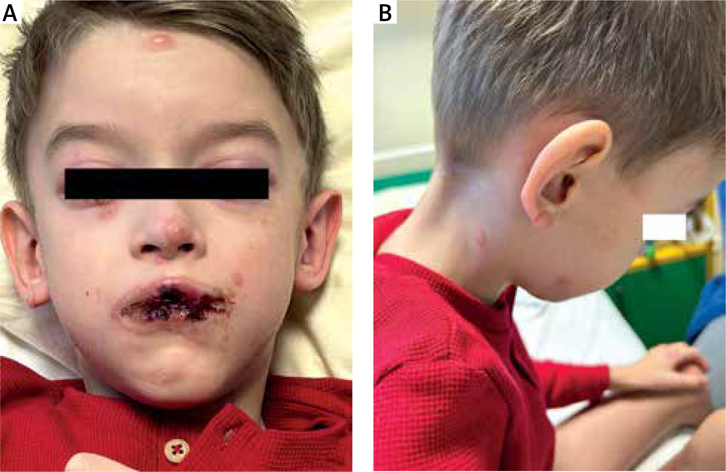
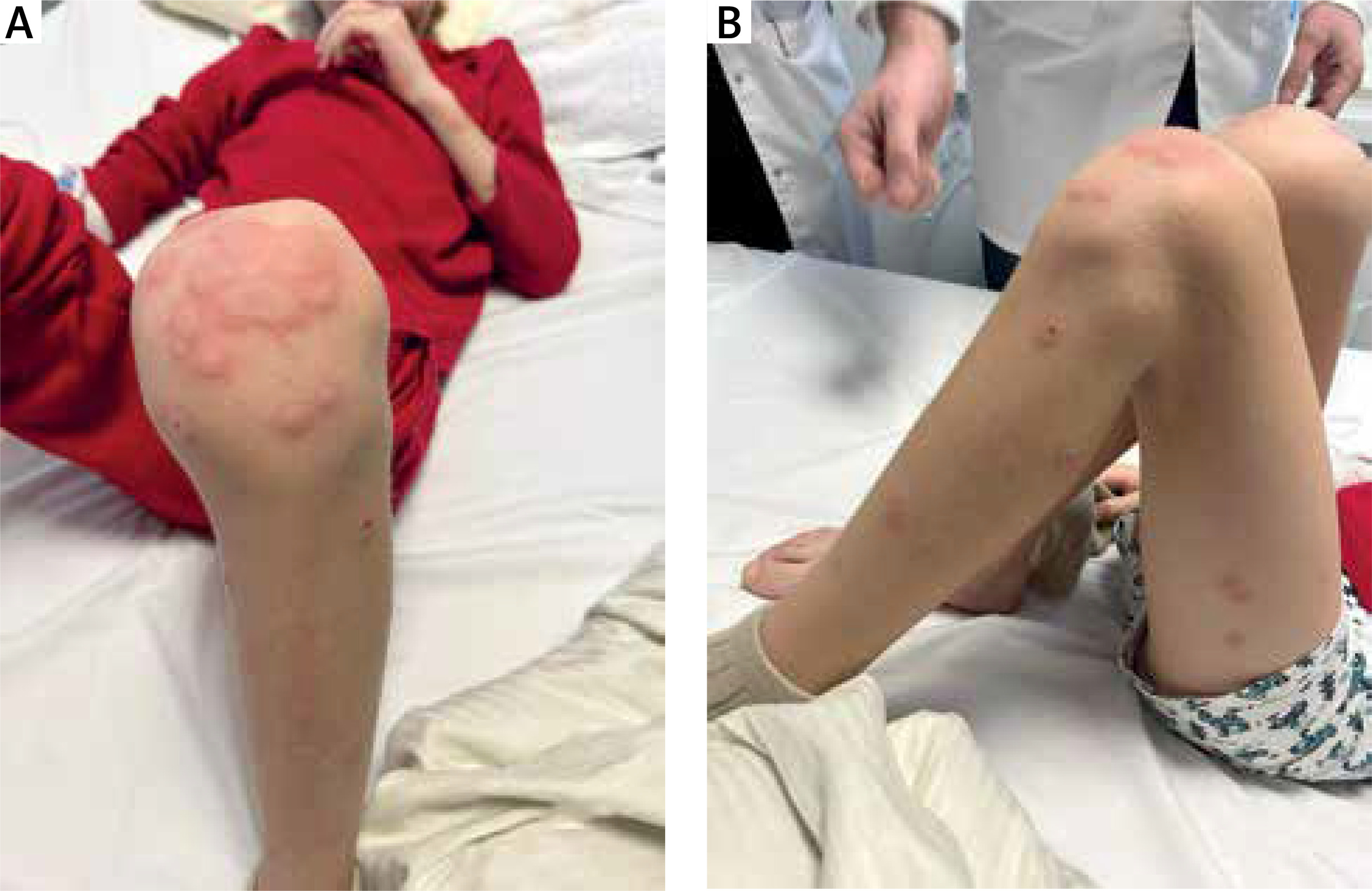
Figure 2
A, B – Patient’s lesions have three concentric zones of colour: a central dark area surrounded by a pale ring, which was surrounded by a red outer ring. The appearance is often described as resembling a “bull’s eye” or “target.” Lesions are often symmetrical and may coalesce
Cyclosporin was gradually tapered over several weeks, and the patient remained lesion-free during the follow-up period. Laboratory parameters were monitored regularly to ensure the safe use of cyclosporin. No significant adverse effects were noted during the treatment course. The skin lesions gradually improved over several weeks, and the child’s overall health returned to normal. Follow-up appointments confirmed resolution of the acute phase of EMM.
Erythema multiforme is a sudden immune-mediated inflammatory skin disorder defined by the presence of raised hives, red spots, and plaques on the skin. It typically appears in a symmetrical pattern on both sides of the body and can also affect the mucous membranes. The skin lesions often have a distinctive target-like appearance. This illness is frequently mistaken for more severe disorders like Stevens-Johnson syndrome (SJS) and toxic epidermal necrosis (TEN). Bastuji-Garin et al. were the first to document EM based on its pathological physiology, causes, and clinical mechanisms [1, 2]. Erythema multiforme is a clinical condition that affects the skin and mucous membranes. It is divided into two categories: erythema multiforme minor and erythema multiforme major, the latter of which is characterized by significant damage to the mucous membranes involving at least two locations [3, 4].
The precise pathobiological mechanisms responsible for the initiation of EM have yet to be completely understood. Infections, medicines, vaccinations, and autoimmune illnesses are considered to be the causative factors for EM. The pathophysiology of EM varies depending on the underlying aetiology. Virus fragments carrying interferon-γ (IFN-γ) to the epithelium increase its expression, leading to the accumulation of T cells in virus-induced EM. IFN-γ subsequently induces the synthesis of cytokines that recruit T and NK cells to the surface of the skin. This is classified as a delayed-type hypersensitivity response. In drug-induced EM, the drug’s metabolites trigger the programmed cell death of keratinocytes, leading to the release of tumor necrosis factor-α (TNF-α) [5]. The HSV is responsible for around 70% of cases of EM, making it the main cause. Mycoplasma pneumoniae (MP) is considered the second most prevalent cause of EM, particularly in youngsters [6, 7]. Furthermore, several medications, including non-steroidal anti-inflammatory drugs, antibiotics such as amoxicillin, norfloxacin, cephalothin, and trimethoprim-sulfamethoxazole, as well as antiepileptic drugs, are commonly linked to EM. Researchers have found a link between the development of EM and barbiturates, phenothiazines, statins, TNF-α inhibitors, and other substances [2, 3, 6].
Particular alleles of the HLA gene have been documented to play a role in the development of more severe skin reactions, such as SJS/TEN and EMM. The HLA-DQB1*03:01 genotype is a prevalent indicator in herpes-associated EM (HAEM) [8].
The prompt identification and management of EM pose a significant obstacle. Controversies arise about the proper classification of EM, SJS, and TEN due to their similarities and overlaps. The classification of these different clinical conditions is based on the specific characteristics of the individual lesions and how they are distributed [9]. SJS, or SJS/TEN, primarily manifests as macules or flat, unusual target lesions and quickly progresses into a blistering condition affecting the skin and mucosal areas [10]. In contrast, EM primarily affects the extremities with target-like lesions, while children often exhibit target-like lesions on the trunk as well.
The diagnostic criteria for SJS and TEN as suggested by Bastuji-Garin et al. [1] in 1993 were presented in Table 1.
Table 1
The diagnostic criteria for SJS and TEN
| Classification | Types of lesions* | Types of lesions* | Percentage of body surface area detached/detachable |
|---|---|---|---|
| Bullous erythema multiforme | Typical or atypical raised targets | Acral | < 10 |
| SJS | Spots ± flat atypical targets | Generalised | < 10 |
| Overlap SJS/TEN | Spots ± flat atypical targets | Generalised | ≥ 10 to 30 |
| TEN with spots | Spots ± flat atypical targets | Generalised | ≥ 30 |
| TEN without spots | No spots or targets | Generalised | ≥ 10 |
Our case report includes the presence of typical target lesions, involvement of the trunk and limbs, extensive mucosal involvement (in at least two sites, specifically the oral and ocular mucous membranes), and systemic symptoms. These all match the diagnostic criteria for EMM, with the skin lesions affecting the whole body surface area (BSA) [9].
Identifying the causative agent for EM is crucial for its treatment. The effectiveness of EM therapy is contingent upon the underlying cause, the severity of the disease, the clinical symptoms, and the progression of EM [10]. The clinical care of EM involves the administration of topical corticosteroids and antihistamines to alleviate symptoms, as well as addressing the root cause of the condition. The administration of systemic corticosteroids has been a common practice for several decades in the treatment of acute EM [11], despite the fact that their specific functions remain uncertain. In cases of drug-induced EM that are extremely suspicious, it is crucial to promptly identify and discontinue the culprit medicines in order to prevent the exacerbation of immunological reactions. Additionally, it is important to avoid any further contact with the same medicine or pharmaceuticals that have similar chemical structures as they may potentially cause cross-reactivity. Cyclosporine works by joining with cyclophilin to make a compound that stops calcineurin from doing its job. Calcineurin is a protein that turns on T lymphocytes. Hence, cyclosporine hampers the activity of T cells and produces an immunosuppressive impact. Cyclosporine has demonstrated efficacy in managing a case of recurrent EM [12], an uncommon and persistent variant of EM that is characterized by its chronic nature and association with the HSV [13]. Additionally, it has been employed in a situation involving severe bullous EM [14] as well as a global bullous fixed drug eruption [15].
No comparative studies have been conducted to assess the efficacy of cyclosporine in comparison to steroids for the treatment of EM. In our case, a paediatric patient exhibited a highly positive clinical outcome when treated with cyclosporine, thereby avoiding the negative side effects associated with corticosteroids.
Erythema multiforme major is a rare but significant dermatological condition in children. Early recognition, appropriate diagnosis, and timely supportive care are crucial for a favourable outcome.